Glaucoma is a disease of the optic nerve, which is the part of the eye that carries the images we see to the brain. The optic nerve is made up of many nerve fibers, like an electric cable containing numerous wires. When pressure inside the eye increases, damage to the optic nerve fibers may occur, causing blind spots to develop. These blind spots usually go undetected until the optic nerve is significantly damaged. If the entire nerve is destroyed, blindness results.
Eyes make fluid (aqueous humor) to nourish its front structures. After nutrients are extracted, the fluid drains away through tiny channels in the front part of the eye called the trabecular meshwork. This fluid balance in the eye creates pressure, intraocular pressure or IOP, which normally ranges from about 10mm to 21mm of mercury. For no apparent reason, the drainage does not working properly, causing the IOP to increase and leading to a gradual damage to the optic nerve. This in turn causes loss of peripheral (side) vision, which gradually progresses towards the central vision.
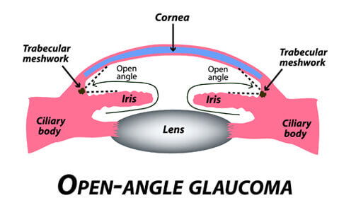
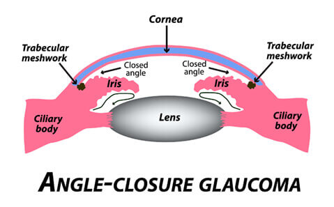
There are two main types of glaucoma, open angle glaucoma and narrow angle glaucoma.
Early detection and treatment by your eye doctor are the keys to preventing optic nerve damage and blindness from glaucoma.
Open angle glaucoma is one in which no cause is identified. There are a few subtypes of open angle glaucoma, but the treatment is the same. People with open angle glaucoma rarely have any symptoms until they have lost a significant amount of side vision.
Narrow angle glaucoma can be acute or chronic. Acute narrow angle glaucoma can cause severe eye pain and sudden damage to the optic nerve. If narrow angles are detected early during routine eye examination, the patient is recommended to have a laser iridotomy to create an opening in the iris (the color part of the eye). This helps to divert the fluid drainage so the acute angle closure would not happen. Chronic narrow angle glaucoma requires laser iridotomy plus eye drops.
Glaucoma is typically detected upon routine eye examination since sufferers do not have symptoms at earlier stages of the disease. During a routine comprehensive eye examination, the eye pressures are taken and the optic nerve is inspected with lens magnification. If eye pressures are high and optic nerves appear to have damage, there is a good chance the patient has glaucoma. Three additional tests are typically done to increase the likelihood of the diagnosis: Pachymetry, HRT/OCT/Disc photos, and visual field examination.
After combining the test results with visual inspection of the nerve, the eye doctor can give a more accurate determination of the presence of glaucoma and its severity.